
Eric will be presenting at MS&T conference
- Post by: siteadmin
- October 1, 2017
- Comments off
Eric Anderson will be presenting his work at Materials Science & Technology 2017, Monday Oct. 9, 10:20 am, room 304.
Additive Manufacturing of Composites and Complex Materials II
Effect of Porosity on Stochastic Fracture of Additive Manufactured Polymer Matrix Composites
Eric Anderson, Ozgur Keles
Consumer level Fused deposition modeling (FDM )systems have been used for production of open-source free designs that contain intentional porosity. Despite the increasing use of FDM, the effect of porosity on the mechanical behavior of FDMed short-carbon-fiber-reinforced composites are unclear. In this talk, we will discuss the effect of internal porosity and extrusion tip vibrations on the mechanical properties and mechanical reliability of the FDMed composites containing up to 50 vol. % porosity. Seven batches of thirty specimens were tensile tested at different porosity levels, a total of ~210 tests. Weibull statistics was used to quantify variations in the mechanical properties. A novel and inexpensive extrusion tip vibration technique was used eliminate road-to-road porosity, which resulted in an increase in both fracture strength and its Weibull modulus from 23 to 54 for dense samples. The resulting microstructures were investigated using X-ray microscopy and scanning electron microscope.
OpenDocument
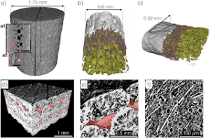
Figure. X-ray computed tomography images of a) as-received filament, b) extruded filament, c) first layer deposition on a hot bed, d) printed composite showing inter-bead porosity (red shade), e) beads with inter- and inner-bead porosiry, and f) fibers and pores at the fiber ends. ABS is shown in gray and fibers are in dark red. Black represents pores in a) and f), and dark green represents pores in b) and c). The porosities (vol. %) are a) 5 , b) 20 , and c) 22. The carbon fiber content (vol. %) are a) 7, b) 6, and c) 10. Scale bars represent 500 μm for a1 and 100 μm for a2. Isotropic voxels of linear dimensions are 1.0 µm for a) and c), 0.83 µm for b), 4.4 µm for d) and e), and 0.9 µm for f). Images were taken by Jeff Gelb at Carl Zeiss X-ray Microscopy.
Posted 1st October 2017 by Ozgur Keles
Categories: Research